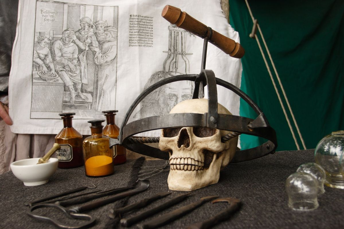
To make his eponymous monster, Victor Frankenstein needed body parts, but organ donation , as we know it, wouldn't emerge for another 135 years or so. And so the fictional doctor "dabbled among the unhallowed damps of the grave" and visited dissecting rooms and slaughterhouses, where he collected parts and pieces like some sort of ghoul.
Les futurs Victor Frankensteins n'auront pas à devenir des pilleurs de tombes pour obtenir des parties du corps. Ils n'auront même pas besoin de corps. Au lieu de cela, nous parions qu'ils tireront parti d'une technologie en développement rapide connue sous le nom de bio -impression . Cette émanation de l'impression 3D vise à permettre aux scientifiques et aux chercheurs médicaux de construire un organe, couche par couche, à l'aide de scanners et d'imprimantes traditionnellement réservés à la conception automobile, à la construction de modèles et au prototypage de produits.
Pour fabriquer un jouet en utilisant cette technique, un fabricant charge une substance, généralement du plastique, dans une machine de la taille d'un mini-réfrigérateur. Il charge également une conception 3D du jouet qu'il veut fabriquer. Lorsqu'il dit à la machine d'imprimer, elle chauffe et, en utilisant le dessin comme un ensemble d'instructions, extrude une couche de plastique fondu à travers une buse sur une plate-forme. Au fur et à mesure que le plastique refroidit, il commence à se solidifier, bien qu'il ne soit rien de plus qu'une seule tranche de l'objet désiré. La plate-forme se déplace alors vers le bas pour qu'une deuxième couche puisse être déposée sur la première. L'imprimante répète ce processus jusqu'à ce qu'elle forme un objet solide ayant la forme du jouet.
Dans les milieux industriels, on parle de fabrication additive car le produit fini est fabriqué en ajoutant de la matière pour créer une forme tridimensionnelle. Elle diffère de la fabrication traditionnelle, qui consiste souvent à soustraire une matière, par usinage, pour obtenir une certaine forme. Les fabricants d'additifs ne se limitent pas à utiliser le plastique comme matière première. Certains utilisent des poudres, qui sont maintenues ensemble par de la colle ou chauffées pour fusionner la poudre. D'autres préfèrent les matières alimentaires, comme le fromage ou le chocolat, pour créer des sculptures comestibles. Et d'autres encore - des versions modernes de Victor Frankenstein - expérimentent des biomatériaux pour imprimer des tissus vivants et, lorsqu'ils sont correctement superposés dans des environnements biotiques, des organes pleinement fonctionnels.
C'est vrai, la même technologie qui peut produire des figurines d'action Star Wars peut également produire des foies, des reins, des oreilles, des vaisseaux sanguins, de la peau et des os humains. Mais imprimer une version 3D de R2-D2 n'est pas exactement la même chose que d'imprimer un cœur qui se dilate et se contracte comme un vrai muscle cardiaque. Coupez une figurine articulée et vous trouverez du plastique de part en part. Coupez à travers un cœur humain et vous trouverez une matrice complexe de cellules et de tissus, qui doivent tous être arrangés correctement pour que l'organe fonctionne. Pour cette raison, la bio-impression se développe plus lentement que les autres techniques de fabrication additive, mais elle progresse. Les chercheurs ont déjà construit des imprimantes 3D modifiées et perfectionnent maintenant les processus qui leur permettront d'imprimer des tissus et des organes pour les tests pharmaceutiques et, finalement, pour la transplantation.
- L'histoire 3D de la bioimpression
- Tout comme une imprimante à jet d'encre, en quelque sorte
- Composants de la bio-imprimante
- Orgues sur mesure
- Un, deux, trois, imprimez !
- Utilisations pour les organes 3D
L'histoire 3D de la bioimpression

La promesse d'imprimer des organes humains a commencé en 1983 lorsque Charles Hull a inventé la stéréolithographie . Ce type spécial d'impression reposait sur un laser pour solidifier un matériau polymère extrudé à partir d'une buse. Les instructions pour la conception provenaient d'un ingénieur, qui définissait la forme 3D d'un objet dans un logiciel de conception assistée par ordinateur (CAO), puis envoyait le fichier à l'imprimeur. Hull et ses collègues ont développé le format de fichier, connu sous le nom de .stl, qui contenait des informations sur la géométrie de la surface de l'objet, représentée sous la forme d'un ensemble de faces triangulaires.
Au début, les matériaux utilisés en stéréolithographie n'étaient pas assez solides pour créer des objets durables. En conséquence, les ingénieurs des premiers jours utilisaient le processus strictement comme un moyen de modéliser un produit final - une pièce de voiture, par exemple - qui serait finalement fabriqué à l'aide de techniques traditionnelles. Toute une industrie, connue sous le nom de prototypage rapide, s'est développée autour de cette technologie et, en 1986, Hull a fondé 3D Systems pour fabriquer des imprimantes 3D et les matériaux nécessaires.
Au début des années 1990, 3D Systems avait commencé à introduire la prochaine génération de matériaux - les nanocomposites , les plastiques mélangés et les métaux en poudre. Ces matériaux étaient plus durables, ce qui signifiait qu'ils pouvaient produire des objets solides et robustes qui pouvaient fonctionner comme des produits finis, et non comme de simples tremplins vers des produits finis.
Il n'a pas fallu longtemps aux chercheurs médicaux pour s'en apercevoir. Qu'est-ce qu'un organe sinon un objet possédant une largeur, une hauteur et une profondeur ? Une telle structure ne pourrait-elle pas être cartographiée en trois dimensions ? Et une imprimante 3D ne pourrait-elle pas recevoir une telle carte et rendre ensuite l'orgue de la même manière qu'elle pourrait rendre un ornement de capot ou un bijou ? Un tel exploit pourrait être facilement accompli si les cartouches d'imprimante pulvérisaient des biomatériaux au lieu de plastiques.
Les scientifiques sont partis à la recherche de tels matériaux et à la fin des années 1990, ils avaient mis au point des techniques et des processus viables pour faire de la construction d'organes une réalité. En 1999, des scientifiques du Wake Forest Institute for Regenerative Medicine ont utilisé une imprimante 3D pour construire un échafaudage synthétique d'une vessie humaine. Ils ont ensuite recouvert l'échafaudage de cellules prélevées sur leurs patients et ont réussi à faire pousser des organes fonctionnels. Cela a ouvert la voie à une véritable bio-impression. En 2002, des scientifiques ont imprimé un rein fonctionnel miniature capable de filtrer le sang et de produire de l'urine dans un modèle animal. Et en 2010, Organovo - une société de bio-impression basée à San Diego - a imprimé le premier vaisseau sanguin.
Aujourd'hui, la révolution continue. Au centre de l'attention se trouvent les imprimantes elles-mêmes, ainsi que le mélange spécial d'encres vivantes qu'elles contiennent. Nous couvrirons les deux ensuite.
Tout comme une imprimante à jet d'encre, en quelque sorte
L'idée de l'impression 3D est directement issue d'une technologie que tout le monde connaît : l' imprimante à jet d'encre . Regardez votre machine HP ou Epson produire une page imprimée et vous remarquerez que la tête d'impression, entraînée par un moteur, se déplace en bandes horizontales sur une feuille de papier. En se déplaçant, l'encre stockée dans une cartouche pulvérise à travers de minuscules buses et tombe sur la page en une série de fines gouttes. Les gouttes s'accumulent pour créer une image, les paramètres de résolution supérieure déposant plus d'encre que les paramètres de résolution inférieure. Pour obtenir une couverture complète de haut en bas, la feuille de papier, située sous la tête d'impression, s'enroule verticalement.
The limitation of inkjet printers is that they only print in two dimensions -- along the x- and y-axes. A 3-D printer overcomes this by adding a mechanism to print along an additional axis, usually labeled the z-axis in mathematical applications. This mechanism is an elevator that moves a platform up and down. With such an arrangement, the ink head can lay down material from side to side, but it can also deposit layers vertically as the elevator draws the platform down and away from the print head. Fill the cartridge with plastic, and the printer will output a three-dimensional plastic widget. Fill it with cells, and it will output a mass of cells.
Conceptually, bioprinting is really that simple. In reality, it's a bit more challenging because an organ contains more than one type of material. And because the material is living tissue, it needs to receive nutrients and oxygen. To accommodate this, bioprinting companies have modified their 3-D printers to better serve the medical community.
Where Can I Find a Bioprinter?
Comme vous pouvez l'imaginer, la technologie de bio-impression n'est pas au point où vous pouvez en commander une sur Amazon, mais vous pouvez trouver, par exemple, la bio-imprimante NovoGen MMX d'Organovo dans des institutions telles que la Harvard Medical School, la Wake Forest University et le Sanford Consortium for Médecine régénérative. Si vous n'êtes pas vraiment un type institutionnel, vous voudrez peut-être consulter le Instructable pour un bioprinter DIY des gens de BioCurious .
Composants de la bio-imprimante

If you were to pull apart a bioprinter, as we'd love to do, you'd encounter these basic parts:
Print head mount -- On a bioprinter, the print heads are attached to a metal plate running along a horizontal track. The x-axis motor propels the metal plate (and the print heads) from side to side, allowing material to be deposited in either horizontal direction.
Elevator -- A metal track running vertically at the back of the machine, the elevator, driven by the z-axis motor, moves the print heads up and down. This makes it possible to stack successive layers of material, one on top of the next.
Platform -- A shelf at the bottom of the machine provides a platform for the organ to rest on during the production process. The platform may support a scaffold, a petri dish or a well plate, which could contain up to 24 small depressions to hold organ tissue samples for pharmaceutical testing. A third motor moves the platform front to back along the y-axis.
Reservoirs -- The reservoirs attach to the print heads and hold the biomaterial to be deposited during the printing process. These are equivalent to the cartridges in your inkjet printer.
Print heads/syringes -- A pump forces material from the reservoirs down through a small nozzle or syringe, which is positioned just above the platform. As the material is extruded, it forms a layer on the platform.
Triangulation sensor -- A small sensor tracks the tip of each print head as it moves along the x-, y- and z-axes. Software communicates with the machine so the precise location of the print heads is known throughout the process.
Microgel -- Unlike the ink you load into your printer at home, bioink is alive, so it needs food, water and oxygen to survive. This nurturing environment is provided by a microgel -- think gelatin enriched with vitamins, proteins and other life-sustaining compounds. Researchers either mix cells with the gel before printing or extrude the cells from one print head, microgel from the other. Either way, the gel helps the cells stay suspended and prevents them from settling and clumping.
Bioink - Les organes sont constitués de tissus et les tissus sont constitués de cellules. Pour imprimer un organe, un scientifique doit pouvoir déposer des cellules spécifiques à l'organe qu'il souhaite construire. Par exemple, pour créer un foie, elle commencerait par des hépatocytes - les cellules essentielles d'un foie - ainsi que d'autres cellules de soutien. Ces cellules forment un matériau spécial appelé bioink , qui est placé dans le réservoir de l'imprimante puis extrudé à travers la tête d'impression. Au fur et à mesure que les cellules s'accumulent sur la plate-forme et s'incrustent dans le microgel, elles prennent une forme tridimensionnelle qui ressemble à un organe humain .
Alternatively, the scientist could start with a bioink consisting of stem cells , which, after the printing process, have the potential to differentiate into the desired target cells. Either way, bioink is simply a medium, and a bioprinter is an output device. Up next, we'll review the steps required to print an organ designed specifically for a single patient.
Made-to-order Organs

When researchers built 3-D printers capable of depositing bioink and forming living masses of cells, they celebrated a major achievement. Then they immediately began to tackle the next big problem: How can bioprinting produce an organ for a specific person? To accomplish this, a medical team needs to collect data about the organ in question -- its size, shape and placement in the patient's body. Then team members need to concoct a bioink using cells taken from the patient. This ensures that the printed organ will be compatible genetically and won't be rejected once it's transplanted in the patient's body.
Pour les organes simples, comme les vessies, les chercheurs n'impriment pas directement les tissus vivants. Au lieu de cela, ils impriment un échafaudage 3D fait de polymères biodégradables ou de collagène. Pour déterminer la forme exacte de l'échafaudage, ils construisent d'abord un modèle 3D à l'aide d'un logiciel de conception assistée par ordinateur (CAO). Ils définissent généralement les coordonnées exactes x, y et z du modèle en prenant des scans du patient à l'aide de la technologie de tomographie informatisée (CT) ou d'imagerie par résonance magnétique (IRM).
Next, researchers get the cells they need by taking a biopsy of the patient's bladder. They then place the cell samples in a culture, where they multiply into a population sufficiently large enough to cover the scaffold, which provides a temporary substrate for the cells to cling to as they organize and strengthen. Seeding the scaffold requires time-consuming and painstaking handwork with a pipette. It generally takes about eight weeks before such artificial bladders are ready for implantation. When doctors finally place the organ in the patient, the scaffold has either disappeared or disappears soon after the surgery.
The procedure above works because bladder tissue only contains two types of cells. Organs like kidneys and livers have a far more complex structure with a greater diversity of cell types. While it would be easy enough to print a scaffold, it would be almost impossible to recreate the three-dimensional structure of the tissue manually. A bioprinter, however, is ideally suited to complete such a time-consuming, detail-oriented task.
One, Two, Three, Print!
Here are the steps to print a complex organ:
- First, doctors make CT or MRI scans of the desired organ.
- Next, they load the images into a computer and build a corresponding 3-D blueprint of the structure using CAD software.
- Combining this 3-D data with histological information collected from years of microscopic analysis of tissues, scientists build a slice-by-slice model of the patient's organ. Each slice accurately reflects how the unique cells and the surrounding cellular matrix fit together in three-dimensional space.
- After that, it's a matter of hitting File > Print, which sends the modeling data to the bioprinter.
- The printer outputs the organ one layer at a time, using bioink and gel to create the complex multicellular tissue and hold it in place.
- Finally, scientists remove the organ from the printer and place it in an incubator, where the cells in the bioink enjoy some warm, quiet downtime to start living and working together. For example, liver cells need to form what biologists call "tight junctions," which describes how the cell membrane of one cell fuses to the cell membrane of the adjacent cell. The time in the incubator really pays off -- a few hours in the warmth turns the bioink into living tissue capable of carrying out liver functions and surviving in a lab for up to 40 days.
The final step of this process -- making printed organ cells behave like native cells -- has been challenging. Some scientists recommend that bioprinting be done with a patient's stem cells. After being deposited in their required three-dimensional space, they would then differentiate into mature cells, with all of the instructions about how to "behave." Then, of course, there's the issue of getting blood to all of the cells in a printed organ. Currently, bioprinting doesn't offer sufficient resolutions to create tiny, single-cell-thick capillaries. But scientists have printed larger blood vessels, and as the technology improves, the next step will be fully functional replacement organs , complete with the vascularization necessary to remain alive and healthy.
Uses for 3-D Organs
At the time of publication, surgeons hadn't implanted an organ printed from scratch into a human. That doesn't mean there haven't been successes. Replacing parts of the skeleton is one area being revolutionized by 3-D printing. Some dentists now take an intra-oral scan of a patient's teeth and send the scan to a lab that fashions a porcelain bridge using a 3-D printer. Prosthetic manufacturers also have changed their approach to designing artificial limbs. Now, many are able to print fairings -- prosthetic limb covers -- that mold perfectly to a person's anatomy, giving the wearer a more comfortable fit. These are just preludes to what the future may hold: printing entire bones for placement in the body. Doctors in the Netherlands have already created a lower mandible on a 3-D printer and implanted the jaw -- made from bioceramic-coated titanium -- in a patient suffering from a chronic bone infection.
Scientists have also successfully printed cartilaginous structures, such as ears and tracheas. To make the former, bioengineers take a 3-D scan of a patient's ear, design a mold using CAD software and then print it out. Then they inject the mold with cartilage cells and collagen. After spending some time in an incubator, the ear comes out, ready for attachment to the patient. A trachea can be made in a similar fashion. In 2012, doctors at the University of Michigan printed a sleeve, made from a 3-D model generated from a CT scan, to wrap and support a baby's trachea, which had been rendered weak and floppy by a rare defect.
The holy grail, of course, is a bioprinted organ, and skin -- the body's largest organ -- may be the first item on the list. Researchers at the Wake Forest Institute for Regenerative Medicine already have developed a complete system to print skin grafts. The system includes a scanner to map a patient's wound and a purpose-built inkjet printer that lays down the cells, proteins and enzymes necessary to form human skin. The goal is to build portable printers for use in field hospitals, where doctors can output skin directly onto patients.
Until these marvels come online, 3-D organs will play an important role in education and drug development. They might even factor into the development of food and clothing products (lab-grown meat and leather). Some medical schools have invested in 3-D printing technology to create surgical models of organs from CT or MRI images. This allows students to practice on hearts, livers and other structures that look and feel just like the real thing. Having access to such lifelike tissues also benefits pharmaceutical companies, which can test candidate drugs to see their effects. Organovo houses several printers capable of printing out three-dimensional models of liver, kidney and cancer tissues. These aren't full organs meant to live indefinitely. Instead, they're "organs on a chip" -- small, biologically active tissue samples designed to respond as native tissues would.
Perhaps one day, bioprinting will make anyone a Victor Frankenstein, capable of printing out organs, bones and muscles and assembling it all into a reasonable facsimile of a human. Then again, there's the issue of a nervous system. Even the best scanners, printers, inks and gels will fall short when it comes to recreating a thinking, dreaming brain. And without that, our efforts would leave us with a collection of anatomically correct, three-dimensionally accurate organs, but nothing to control them.
Who's the Boss of Bioprinting?
In the U.S., that role would fall to the Food and Drug Administration, but this is new territory for the agency, too. According to an Aug. 15, 2013, blog post, two labs in the agency's Office of Science and Engineering Laboratories (OSEL) are on the case. The Laboratory for Solid Mechanics is busy evaluating "how different printing techniques and processes affect the strength and durability of the materials used in medical devices." The Functional Performance and Device Use Laboratory has "developed and adapted computer-modeling methods to help us determine the effect of design changes on the safety and performance of devices when used in different patient populations" [source: Pollack and Coburn].
Lots More Information
Author's Note: How 3-D Bioprinting Works
I remember my first printer: a Brother typewriter hooked to a Commodore 64, followed by a daisy-wheel printer powered by an IBM PC. Hard to believe we might have bioprinters sitting on our desktops one day. If we do, I wonder where we'll go to get new bioink cartridges?
Related Articles
- How 3-D Printing Works
- How Stereolithography 3-D Layering Works
- How Inkjet Printers Work
- How Artificial Hearts Work
- How can scientists use an inkjet printer to make bones?
- How Organ Donation Works
- How Organ Transplants Work
Sources
- Atala, Anthony. "Printing a human kidney." TED Talks. March 2011. (Nov. 17, 2013)http://www.ted.com/talks/anthony_atala_printing_a_human_kidney.html
- Banham, Russ. "Printing a Medical Revolution." T. Rowe Price. May 2012. (Nov 17, 2013)http://individual.troweprice.com/public/Retail/Planning-&-Research/Connections/3D-Printing/Printing-a-Medical-Revolution
- Clark, Liat. "Bioengineer: the heart is one of the easiest organs to bioprint, we'll do it in a decade." Cardiovascular Innovation Institute. Nov. 21, 2013. (Nov. 25, 2013)http://cv2i.org/bioengineer-heart-one-easiest-organs-bioprint-well-decade/
- Dutton, Gail. "3D Printing May Revolutionize Drug R&D." Genetic Engineering & Biotechnology News. Nov. 15, 2013. (Nov. 17, 2013)http://www.genengnews.com/gen-articles/3d-printing-may-revolutionize-drug-r-d/5062/
- Fountain, Henry. "At the Printer, Living Tissue." The New York Times. Aug. 18, 2013. (Nov. 17, 2013) http://www.nytimes.com/2013/08/20/science/next-out-of-the-printer-living-tissue.html?
- History.com Staff. "Organ Transplants: A Brief History." History.com. Feb. 21, 2012. (Nov. 17, 2013) http://www.history.com/news/organ-transplants-a-brief-history
- Hsu, Jeremy. "3D Printed Organs May Mean End To Waiting Lists, Deadly Shortages." Huffington Post. Sept. 25, 2013. (Nov. 17, 2013)http://www.huffingtonpost.com/2013/09/25/3d-printed-organs_n_3983971.html
- Hsu, Jeremy. "3D Printing: What a 3D Printer Is and How It Works." LiveScience. May 21, 2013. (Nov. 17, 2013) http://www.livescience.com/34551-3d-printing.html
- Hsu, Jeremy. "Tiny 3D-Printed Organs Aim for 'Body on a Chip'." LiveScience. Sept. 16, 2013. (Nov. 17, 2013) http://www.livescience.com/39660-3d-printed-body-on-a-chip.html
- Image Specialists. "How Inkjet Printers Work."http://www.image-specialists.com/ink_int_injet_printer.aspx
- Leckart, Steven. "The Body Shop, Popular Science." Popular Science. August 2013.
- Organovo. "The Bioprinting Process." (Nov. 17, 2013)http://www.organovo.com/science-technology/bioprinting-process
- Pollack, Steven K. and Coburn, James. "FDA goes 3-D." FDAVoice. Aug. 15, 2013. (Dec. 13, 2013) http://blogs.fda.gov/fdavoice/index.php/2013/08/fda-goes-3-d/
- Royte, Elisabeth. "Qu'est-ce qui nous attend pour l'impression 3D ?" Magazine Smithsonien. Mai 2013. (17 novembre 2013) http://www.smithsonianmag.com/science-nature/What-Lies-Ahead-for-3-D-Printing-204136931.html
- Ungar, Laura. "Les chercheurs se rapprochent de l'impression de cœurs en 3D." États-Unis aujourd'hui. 29 mai 2013. (17 novembre 2013) http://www.usatoday.com/story/tech/2013/05/29/health-3d-printing-organ-transplant/2370079/
- Wake Forest Institute pour la médecine régénérative. "Utilisation de la technologie à jet d'encre pour imprimer des organes et des tissus." 2 août 2013. (17 novembre 2013) http://www.wakehealth.edu/Research/WFIRM/Our-Story/Inside-the-Lab/Bioprinting.htm